

-
Laser
-
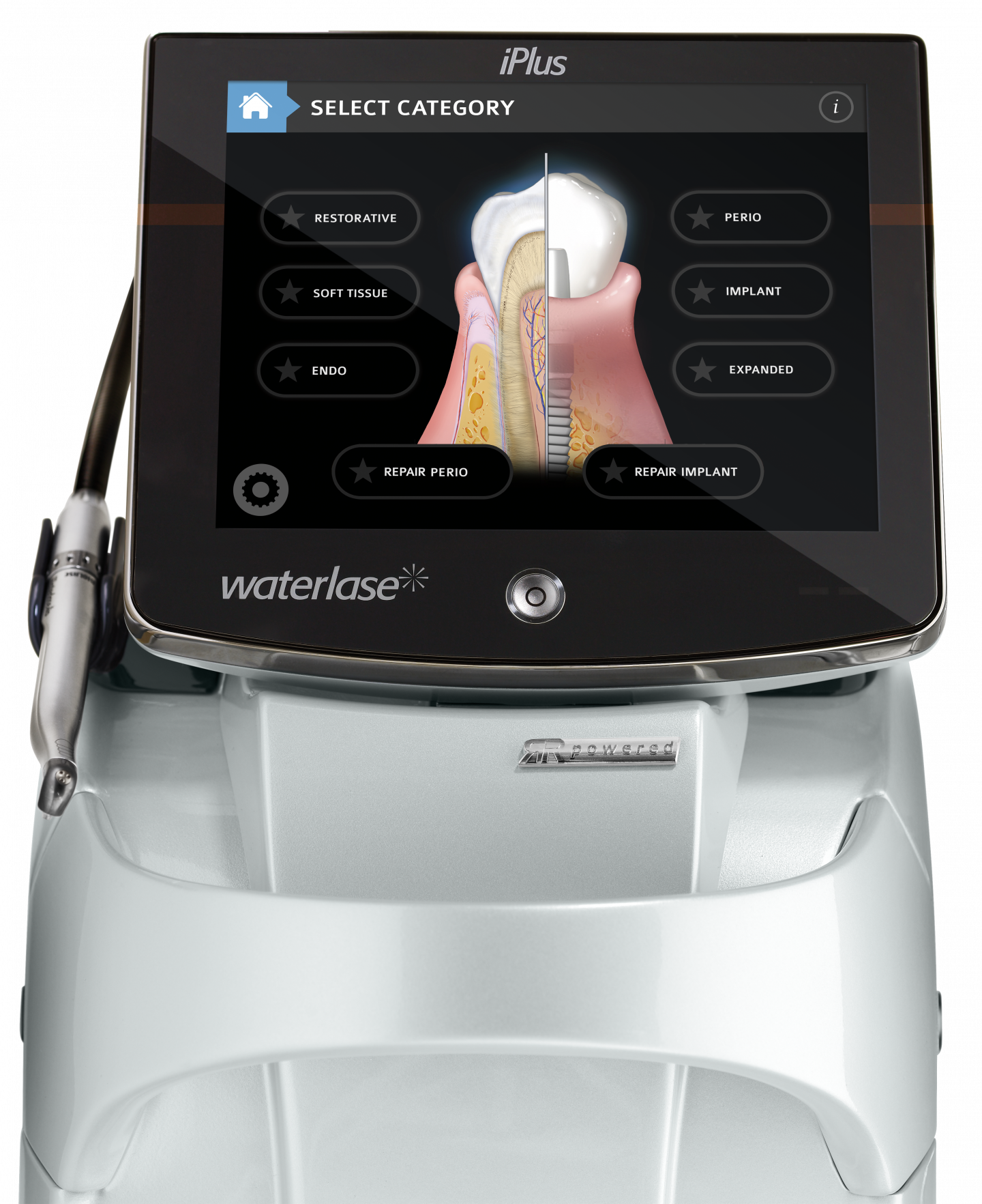
Waterlase iPlusTM - intuitiv und vielseitig
Der Waterlase iPlusTM ist der vielseitigste Dental-Laser auf dem Markt. Dank der einzigartigen Lasertechnologie eignet er sich zur Behandlung von Weich- und Hartgewebe, einschließlich Knochen.
-
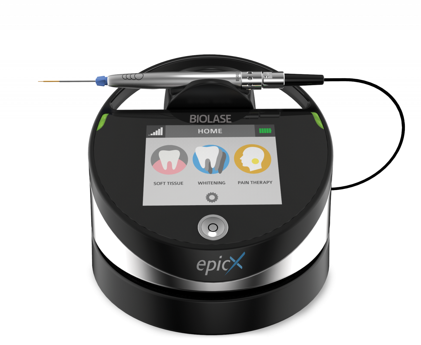
Epic XTM Diodenlaser
Mit dem kleinen, tragbaren Epic XTM Dentallaser schneiden Sie Weichgewebe schonend, schnell und blutungsarm; setzen ihn zur Desinfektion in der Endodontie und Parodontologie ein, stoppen Blutungen, entfernen Hämangiome, lindern Schmerzen, helfen bei Aphten und Herpes und beschleunigen die Bleaching Behandlung.
-
Epic QTM Diodenlaser
Der Epic QTM vereint Langlebigkeit mit Zuverlässigkeit und bietet die neueste
Technologie zur Behandlung von Weichgewebe
in einem tragbaren, erschwinglichen Paket.
-
- Klinische Studien
- Downloads
- Kontakt
- Shop

Klinische Studien zur Implantologie mit dem Dental-Laser
| Groups | Link | Author | Title | Journal | Year | Rating |
|---|---|---|---|---|---|---|
| Implant | URL | Abd El daym, D.A., Gheith, M.E., Abbas, N.A., Rashed, L.A. and Abd El Aziz, Z.A. | Biocompatibility of erbium chromium-doped yattrium-scandium-gallium-garnet ( Er , Cr : YSGG 2780 nm ) laser-treated titanium alloy used for dental applications ( in vitro study ) [Abstract] |
Lasers in Dental Science Vol. April, pp. 1-6 |
2018 | rank2 |
| Abstract: Abstract The use of dental implants in the partial and complete edentulism has become the primary treatment regimen in themodern dentistry. Erbiumchromium-doped yattrium-scandium-gallium-garnet (Er,Cr:YSGG) laser is most often used in dentistry in implant surgery and management of peri-implantitis. The aim of the present study was to assess the biocompatibility of Er,Cr:YSGGlaser-treated titanium alloy (Ti-6Al-4V) and its surface characteristics to understand the impact of the laser on the titanium alloy surfaces. Materials and methods A total of 20 discs of titanium alloy (Ti-6Al-4V) were used. Ten discs were irradiated with Er,Cr:YSGG laser which was operating in a normal room atmosphere and temperature at power 2 W. Biocompatibility was investigated in vitro via MTT assay. Surface analysis of laser-treated and laser-untreated discs was examined with a scanning electron microscope. Result Laser-treated group showed superior cell viability compared to untreated group. No undesirable changes were observed by SEM. Conclusion We can conclude that Er,Cr:YSGG laser safely could improve the biocompatibility of dental implant. | ||||||
| Implant | URL | Abd El daym, D.A., Gheith, M.E., Abbas, N.A., Rashed, L.A. and Abd El Aziz, Z.A. | Corrosion behavior of erbium chromium-doped yattrium-scandium-gallium-garnet (Er,Cr:YSGG 2780 nm) laser-treated titanium alloy used for dental applications at different pH conditions (in vitro study) [Abstract] |
Lasers in Dental Science Vol. April, pp. 1-10 |
2018 | rank2 |
| Abstract: Abstract The use of dental implants in the partial and complete edentulism has become the primary treatment regimen in themodern dentistry. Erbiumchromium-doped yattrium-scandium-gallium-garnet (Er,Cr:YSGG) laser is most often used in dentistry in implant surgery and management of peri-implantitis. The aim of the present study was to assess the biocompatibility of Er,Cr:YSGGlaser-treated titanium alloy (Ti-6Al-4V) and its surface characteristics to understand the impact of the laser on the titanium alloy surfaces. Materials and methods A total of 20 discs of titanium alloy (Ti-6Al-4V) were used. Ten discs were irradiated with Er,Cr:YSGG laser which was operating in a normal room atmosphere and temperature at power 2 W. Biocompatibility was investigated in vitro via MTT assay. Surface analysis of laser-treated and laser-untreated discs was examined with a scanning electron microscope. Result Laser-treated group showed superior cell viability compared to untreated group. No undesirable changes were observed by SEM. Conclusion We can conclude that Er,Cr:YSGG laser safely could improve the biocompatibility of dental implant. | ||||||
| Repair Implant | DOI URL | Al-Falaki, R., Cronshaw, M. and Hughes, F.J. | Treatment outcome following use of the erbium, chromium:yttrium, scandium, gallium, garnet laser in the non-surgical management of peri-implantitis: a case series. [Abstract] |
British dental journal Vol. 217(8), pp. 453-457 |
2014 | rank5 |
| Abstract: AIM: To date there is no consensus on the appropriate usage of lasers in the management of peri-implantitis. Our aim was to conduct a retrospective clinical analysis of a case series of implants treated using an erbium, chromium:yttrium, scandium, gallium, garnet laser. MATERIALS AND METHODS: Twenty-eight implants with peri-implantitis in 11 patients were treated with an Er,Cr:YSGG laser (68 sites >4 mm), using a 14 mm, 500 mum diameter, 60 degrees (85%) radial firing tip (1.5 W, 30 Hz, short (140 mus) pulse, 50 mJ/pulse, 50% water, 40% air). Probing depths were recorded at baseline after 2 months and 6 months, along with the presence of bleeding on probing. RESULTS: The age range was 27-69 years (mean 55.9); mean pocket depth at baseline was 6.64 +/- SD 1.48 mm (range 5-12 mm),with a mean residual depth of 3.29 +/- 1.02 mm (range 1-6 mm) after 2 months, and 2.97 +/- 0.7 mm (range 1-9 mm) at 6 months. Reductions from baseline to both 2 and 6 months were highly statistically significant (P <0.001). Patient level reduction in bleeding from baseline to both 2 and 6 months were statistically significant (P <0.001). CONCLUSION: In view of the positive findings in this pilot study, well-designed randomised controlled trials of the use of Er,Cr:YSGG laser in the non-surgical management of peri-implantitis are required to validate our clinical findings. | ||||||
| Implant | DOI | Aoki, A., Mizutani, K., Schwarz, F., Sculean, A., Yukna, R., Takasaki, A., Romanos, G., Taniguchi, Y., Sasaki, K., Zeredo, J., Koshy, G., Coluzzi, D., White, J., Abiko, Y., Ishikawa, I. and Izumi, Y. | Periodontal and peri-implant wound healing following laser therapy [Abstract] |
Periodontology 2000 Vol. 68, pp. 217-269 |
2015 | rank5 |
| Abstract: Laser irradiation has numerous favorable characteristics, such as ablation or vaporization, hemostasis, biostimulation (photobiomodulation) and microbial inhibition and destruction, which induce various beneficial therapeutic effects and biological responses. Therefore, the use of lasers is considered effective and suitable for treating a variety of inflammatory and infectious oral conditions. The CO2, neodymium-doped yttrium-aluminium-garnet (Nd:YAG) and diode lasers have mainly been used for periodontal soft-tissue management. With development of the erbium-doped yttrium-aluminium-garnet (Er:YAG) and erbium, chromium-doped yttrium-scandium-gallium-garnet (Er,Cr:YSGG) lasers, which can be applied not only on soft tissues but also on dental hard tissues, the application of lasers dramatically expanded from periodontal soft-tissue management to hard-tissue treatment. Currently, various periodontal tissues (such as gingiva, tooth roots and bone tissue), as well as titanium implant surfaces, can be treated with lasers, and a variety of dental laser systems are being employed for the management of periodontal and peri-implant diseases. In periodontics, mechanical therapy has conventionally been the mainstream of treatment; however, complete bacterial eradication and/or optimal wound healing may not be necessarily achieved with conventional mechanical therapy alone. Consequently, in addition to chemotherapy consisting of antibiotics and anti-inflammatory agents, phototherapy using lasers and light-emitting diodes has been gradually integrated with mechanical therapy to enhance subsequent wound healing by achieving thorough debridement, decontamination and tissue stimulation. With increasing evidence of benefits, therapies with low- and high-level lasers play an important role in wound healing/tissue regeneration in the treatment of periodontal and peri-implant diseases. This article discusses the outcomes of laser therapy in soft-tissue management, periodontal nonsurgical and surgical treatment, osseous surgery and peri-implant treatment, focusing on postoperative wound healing of periodontal and peri-implant tissues, based on scientific evidence from currently available basic and clinical studies, as well as on case reports. | ||||||
| Implant | DOI URL | Arnabat-Domínguez, J., Bragado-Novel, M., España-Tost, A.J., Berini-Aytés, L. and Gay-Escoda, C. | Advantages and esthetic results of ErCrYSGG laser application in second-stage implant surgery in patients with insufficient gingival attachment: a report of three cases. [Abstract] |
Lasers in medical science Vol. 25(3), pp. 459-64 |
2010 | rank5 |
| Abstract: Traditional implant placement involves two surgical stages. Although the second stage is comparatively less aggressive for the patient, postoperative pain and swelling can be further reduced by the use of laser instead of a scalpel. Correct handling of peri-implant soft tissue is of major importance in obtaining adequate gingival tissue attachment around implants. The presence of this keratinized gingiva ensures adequate esthetic results and maintains implant health. We report on three patients with implants in the anterior area who were operated on under the above conditions. Traditionally, the tissue overlying the implants is removed and eliminated. In seeking a way to preserve the attached gingiva, we raised a trapezoidal flap, uncovering each implant and allowing apical repositioning and transpositioning of keratinized gingiva to the buccal side. The results obtained were compared with those from other patients operated on by conventional scalpel. The erbium, chromium:yttrium-scandium-gallium-garnet (Er,Cr:YSGG) laser minimized postoperative pain, and the time to prosthetic rehabilitation was also shortened. The esthetic results were far superior, and no complications were recorded. | ||||||
| Implant | URL | Berk, G., Franzen, R., Atici, K., Hakki, S.S., Berk, N. and Gutknecht, N. | Evaluation of contaminated implant surfaces irradiated by ErCrYSGG Laser [Abstract] |
Journal of Implant and Advanced Clinical Dentistry Vol. 5(4), pp. 19-26 |
2013 | |
| Abstract: Background: The aim of the study was to evaluate the efficiency of an Er,Cr:YSGG laser to remove the biofilm of the contaminated implant surfaces and to examine the possible alterations on the surface structure after laser irradiation. Methods: Nine failed SLA surfaced dental implants were used for laser irradiation and one unused implant served as control. Of the nine contaminated implants, eight were marked vertically to create two sides on the same implant, and the ninth contaminated implant was kept as a control implant of the failed implant group. One side of the contaminated implants was treated with Er,Cr:YSGG laser while the other side remained untreated. The surfaces were irradiated with a pulsed Er,Cr:YSGG laser with a power setting of 1.5 W, 15 Hz, 140 μs pulse duration for 30 seconds per thread in the non-contact mode. After macroscopic picture evaluation, light microscopic evaluation and SEM analysis were performed subsequently. Results: SEM and light microscopy images demonstrated that Er,Cr:YSGG laser treatment with predetermined settings for titanium surfaces was able to remove all the contaminants and debris from the implant surfaces without any surface alteration which was also noticeable in the macroscopic pictures. Conclusion: It was concluded that the use of Er,Cr:YSGG laser with current settings was efficient to decontaminate implant surfaces and the results of this study suggested that Er,Cr:YSGG laser can be used safely in the clinics for the treatment of peri-implantitis for biofilm removal without damaging the implant surface. | ||||||
| Implant | URL | Cassoni, A., Miranda, P.V., Rodrigues, J.A., Heluy, S.C.d.L., Blay, A. and Shibli, J.A. | Thermal effects on zirconia substrate aſter Er,Cr:YSGG irradiation [Abstract] |
Revista de Odontologia da UNESP Vol. 42(6), pp. 439-443 |
2013 | rank4 |
| Abstract: Objective: The objective of the present study was to investigate the thermal effects of Er,Cr:YSGG laser irradiation (1.5W/20Hz) on yttrium-stabilized tetragonal zirconia polycrystal (Y-TZP). Material and method: Fifteen disks of Y-TZP (AS Technology TitaniumFIX, São José dos Campos, Brazil) with 5 mm diameter and 3 mm high standardized with CAD-CAM were used. The Y-TZP disks were randomized in three groups (n=5): Y-TZP-G1 = control (no laser treatment); Y-TZP-G2 = Y-TZP + Er,Cr:YSGG laser (air-water cooling proportion 80%/25%); Y-TZP-G3 = Y-TZP + Er,Cr:YSGG laser (air-water cooling proportion 80%/0%). A thermopar (SmartMether, Novus, Porto Alegre, RS, Brazil) was attached to a digital thermometer (SmartMether, Novus, Porto Alegre, RS, Brazil) fixed to the opposite irradiated surface. The temperature gradients (∆T) were calculated (∆T = Final Temperature – Initial Temperature) for each group. Values were statistically analyzed by one-way ANOVA at the 95% confidence level and compared by Tukey post-hoc test (α=0.05) for each material. One sample of each group was analyzed by confocal white light microscopy. Result: The ANOVA test showed significant differences for the factor “laser” (p<.001). The temperature gradients (∆T value) showed the following results: Y-TZP-G1 = 0 °C; Y-TZP-G2 = –1.4 °C and Y-TZP-G3 = 21.4 °C. The ∆T values (°C) of the non-refrigerated group were higher than the refrigerated group. The roughness value (Ra) ranged from 4.50 to –33.65 µm. Conclusion: The water refrigeration for Er,Cr:YSGG irradiation is essential to avoid thermal increase in the Y-TZP | ||||||
| Implant | DOI | Ercan, E., Arin, T., Kara, L., Çandirli, C. and Uysal, C. | Effects of Er,Cr:YSGG laser irradiation on the surface characteristics of titanium discs: An in vitro study [Abstract] |
Lasers in Medical Science Vol. 29(3), pp. 875-880 |
2014 | rank1 |
| Abstract: Lasers are used to modify the surfaces of dental implants or to decontaminate exposed implant surfaces. However, research is lacking on whether the laser causes any change on the surfaces of titanium implants. We aimed to determine the effects of laser treatment on the surface characteristics of titanium discs. Nine discs were fabricated using grade-V titanium with resorbable blast texturing surface characteristics. The discs were irradiated with an erbium, chromium: yttrium, scandium, gallium, garnet (Er,Cr:YSGG) laser under different experimental conditions (R1-9). Scanning electron microscopy was used to evaluate implant surface topography qualitatively, and a mechanical contact profilometer was used to evaluate surface roughness. The R3 and R5 parameters caused no measurable change. Minor cracks and grooves were observed in discs treated with the R1, R2, R4, R7 and R9 parameters. Major changes, such as melting, flattening and deep crack formation, were observed in discs subjected to R6 (2 W, 30 Hz, 2 mm. distance, 30 s) and R8 (3 W, 25 Hz, 2 mm. distance, 45 s) parameters. The lowest surface roughness value was obtained with the R8 parameter. Irradiation distance, duration, frequency and power were the most significant factors affecting surface roughness. Parameters such as wavelength, output power, energy, dose and duration should be considered during irradiation. The results of this study indicate that the distance between the laser tip and the irradiated surface should also be considered. | ||||||
| Implant | DOI URL | Gomez-Santos, L., Arnabat-Dominguez, J., Sierra-Rebolledo, a. and Gay-Escoda, C. | Thermal increment due to ErCr:YSGG and CO2 laser irradiation of different implant surfaces. A pilot study [Abstract] |
Medicina Oral, Patologia Oral y Cirugia Bucal Vol. 15(5), pp. e782-e787 |
2010 | rank5 |
| Abstract: OBJECTIVE: An evaluation and comparison is made of the thermal increment at different implant surfaces during irradiation with CO2 and ErCr:YSGG lasers.nnSTUDY DESIGN: Five threaded and impacted implants with four types of surfaces were inserted in an adult pig rib: two implants with a hydroxyapatite surface (HA) (impacted and threaded, respectively), a machined titanium surface implant (TI mach), a titanium plasma spray surface implant (TPS), and a sandblasted, acid-etched surface implant (SBAE). A 0.5-mm diameter bone defect was made in the implant apical zone, and a type-K thermocouple (Termopar) was placed in contact with the implant. The implants were irradiated in the coronal zone of each implant with a CO2 (4 W continuous mode) and an ErCr:YSGG laser (1.5 W, pulsed mode) first without and then with refrigeration. The temperature variations at the implant apical surface were recorded.n An apical temperature increase was recorded in all cases during CO2 and ErCr:YSGG laser irradiation without refrigeration. However, when the ErCr:YSGG was used with a water spray, a decrease in temperature was observed in all implants. The acid-etched and sandblasted surfaces were those most affected by the thermal changes.n The ErCr:YSGG laser with a water spray applied to the sealing cap or coronal zone of the implants does not generate thermal increments in the apical surface capable of adversely affecting osseointegration and the integrity of the peri-implant bone tissue. | ||||||
| Implant | URL | Kusek, E.R. | The Er,Cr: YSGG Laser : A Perfect Fit with Implant Dentistry | Journal of Laser Dentistry Vol. 18(3), pp. 132-134 |
2010 | rank4 |
| Socket Debridement | DOI URL | Lee, S.-Y., Piao, C., Heo, S.-J., Koak, J.-Y., Lee, J.-H., Kim, T.-H., Kim, M.-J., Kwon, H.-B. and Kim, S.-K. | A comparison of bone bed preparation with laser and conventional drill on the relationship between implant stability quotient (ISQ) values and implant insertion variables. [Abstract] |
The journal of advanced prosthodontics Vol. 2(4), pp. 148-53 |
2010 | rank3 |
| Abstract: PURPOSE: The aim of this study was to investigate a comparison of implant bone bed preparation with Er,Cr:YSGG laser and conventional drills on the relationship between implant stability quotient (ISQ) values and implant insertion variables.nnMATERIALS AND METHODS: Forty implants were inserted into two different types of pig rib bone. One group was prepared with conventional drills and a total of 20 implants were inserted into type I and type II bone. The other group was prepared with a Er,Cr:YSGG laser and a total of 20 implants were inserted into type I and type II bone. ISQ, maximum insertion torque, angular momentum, and insertion torque energy values were measured.n The mean values for variables were significantly higher in type I bone than in type II bone (P < .01). In type I bone, the ISQ values in the drill group were significantly higher than in the laser group (P < .05). In type II bone, the ISQ values in the laser group were significantly higher than in the drill group (P < .01). In both type I and type II bone, the maximum insertion torque, total energy, and total angular momentum values between the drill and laser groups did not differ significantly (P ≥ .05). The ISQ values were correlated with maximum insertion torque (P < .01, r = .731), total energy (P < .01, r = .696), and angular momentum (P < .01, r = .696).n Within the limitations of this study, the effects of bone bed preparation with Er,Cr:YSGG laser on the relationship between implant stability quotient (ISQ) values and implant insertion variables were comparable to those of drilling. | ||||||
| Implant | DOI | Mellado-Valero, A., Buitrago-Vera, P., Sol??-Ruiz, M.F. and Ferrer-Garc??a, J.C. | Decontamination of dental implant surface in peri-implantitis treatment: A literature review [Abstract] |
Medicina Oral, Patologia Oral y Cirugia Bucal Vol. 18(6) |
2013 | rank4 |
| Abstract: OBJECTIVE: The aim of this human in vivo pilot study was to evaluate the efficacy of six antimicrobial agents on the surface decontamination of an oral biofilm attached to titanium implants.n For in vivo biofilm formation, we fixed titanium specimens to individual removable acrylic upper jaw splints (14 specimens in every splint), which were worn by four volunteers overnight for 12 h. The specimens were then treated with different antimicrobial agents for 1 min (Sodium hypochlorite, Hydrogen peroxide 3%, Chlorhexidingluconate 0.2%, Plax, Listerine, citric acid 40%). Afterwards, we quantified the total bacterial load and the viability of adhering bacteria by live or dead cell labelling in combination with fluorescence microscopy.n The total bacterial load on the titanium surfaces was significantly higher after incubation in the control solution phosphate-buffered saline (PBS) than after disinfection in sodium hypochlorite, hydrogen peroxide, chlorhexidine, Plax, Listerine, and citric acid. Furthermore, a significantly lower ratio between dead and total adhering bacteria (bactericidal effect) was found after incubation in control PBS, Plax mouth rinse, and citric acid than after incubation in sodium hypochlorite, hydrogen peroxide, chlorhexidine, and Listerine.n All tested antiseptics seem to be able to reduce the total amount of microorganisms accumulating on titanium surfaces. Furthermore, sodium hypochlorite, hydrogen peroxide, chlorhexidine, and Listerine showed a significant bactericidal effect against adhering bacteria. | ||||||
| Implant | DOI URL | Miller, R.J. | Treatment of the contaminated implant surface using the Er,Cr:YSGG laser. [Abstract] |
Implant dentistry Vol. 13, pp. 165-170 |
2004 | rank5 |
| Abstract: Treatment of the contaminated implant surface by mechanical and chemotherapeutic means has met with mixed success. Incomplete surface debridement or alteration of the implant surface could compromise attempts at grafting and reintegration of the implant body. Development of a laser system operating at 2780 nm and using an ablative hydrokinetic process offers the possibility for more efficient decontamination and debridement. The Er,Cr: YSGG laser is evaluated and compared with the most commonly used chemotherapeutic modality for treatment of the implant surface. A scanning electron microscope study is presented comparing YSGG ablation to citric acid treatment of the titanium plasma sprayed and HA-coated implant surface. We can conclude that laser ablation using the YSGG laser is highly efficient at removing potential contaminants on the roughened implant surface while demonstrating no effects on the titanium substrate. | ||||||
| Socket Debridement | DOI | Montoya-Salazar, V., Castillo-Oyague, R., Torres-Sanchez, C., Lynch, C.D., Gutierrez-Perez, J.L. and Torres-Lagares, D. | Outcome of single immediate implants placed in post-extraction infected and non-infected sites, restored with cemented crowns: A 3-year prospective study [Abstract] |
Journal of Dentistry Vol. 42(6), pp. 645-652 |
2014 | rank3 |
| Abstract: Objectives To compare the survival of immediate implants placed in postextraction infected and non-infected sites, restored with cemented crowns. Methods Thirty-six implants were immediately placed in non-infected sockets (control group (CG), n = 18), and in infected alveoli (test group (TG), n = 18) that had been debrided, curetted, cleaned with 90% hydrogen peroxide, irradiated with yttrium-scandium-gallium-garnet (Er,Cr:YSGG) laser, and irrigated with a sterile solution. Guided bone regeneration was performed under antibiotic coverture. All study patients had both a CG and a TG site. The implant osteotomy sites were extended 3-4 mm beyond the apical extent of the sockets to achieve primary stability for the implants. The prosthetic phase occurred 4.5 months after surgery. Success criteria were accepted as the presence of implant stability, absence of a radiolucent zone around the implants, absence of mucosal suppuration, and lack of pain. Clinical evaluations were performed at baseline, and at 12, 24, and 36 months of follow-up. Results All of the implants were osseointegrated 3 months after surgery. The 3-year survival rate was 94.44% for TG, and 100% for CG. The clinical and radiographic variables tested yielded no significant differences among groups at 36 months. Conclusions Under the tested conditions, immediate implant placement can be considered a predictable treatment option for the restoration of fresh postextraction infected sockets. Clinical significance Immediate implants may be indicated for replacing teeth lost due to chronic periapical lesions with endodontic failure history when appropriate preoperative procedures are taken to clean and decontaminate the surgical sites. ?? 2014 Elsevier Ltd. All rights reserved. | ||||||
| Implant | URL | Smeo, K., Nasher, R. and Gutknecht, N. | Antibacterial effect of Er , Cr : YSGG laser in the treatment of peri-implantitis and their effect on implant surfaces : a literature review [Abstract] |
Lasers in Dental Science Vol. April, pp. 1-9 |
2018 | rank4 |
| Abstract: The aim Thepresent study aimsto conduct adescriptive analysisbyreviewing in vivo and invitro studies concernedwith the antibacterial effect of Er,Cr:YSGG laser (2780 nm) and their effects on implant surfaces at different parameters for peri-implantitis treatment. Materials and methods The PubMed and Google Scholar had been used to search for articles that focused on the antibacterial effect of Er,Cr:YSGG laser (2780 nm) in the treatment of peri-implantitis and their effects on implant surfaces. This literature search was limited to ten years (January 2007–March 2017). Results The favorable settings of Er,Cr:YSGG laser which can be used significantly to treat peri-implantitis without titanium surface characteristic alteration or increase of temperature based on reviewed articles are in a non-contactmode (0.5mmdistance) at 1 and 1.5W, 10 and 30 Hz, and 60 and 120 s, respectively, under air and water flow for refrigeration. Conclusion Er,Cr:YSGG laser with specific parameters is an effective tool in the treatment of peri-implantitis without negative effect on the implant surfaces | ||||||
| Implant | DOI | Smith, L.P. and Rose, T. | Laser explantation of a failing endosseous dental implant [Abstract] |
Australian Dental Journal Vol. 55(2), pp. 219-222 |
2010 | rank3 |
| Abstract: Explantation of failed dental implants has traditionally been performed by mechanical bone removal techniques. The advent of intraoral laser surgery has seen increasing numbers of applications in oral implantology. The technique demonstrates safe and efficient explantation of a failed dental implant using Er,Cr:YSGG laser. Laser assisted explantation of dental implants is a minimally invasive technique providing an alternative to conventional mechanical explantation techniques. | ||||||
| Implant | DOI URL | Strever, J.M., Lee, J., Ealick, W., Peacock, M., Shelby, D., Susin, C., Mettenberg, D., El-Awady, A., Rueggeberg, F. and Cutler, C.W. | Erbium, Chromium:Yttrium-Scandium-Gallium-Garnet Laser Effectively Ablates Single-Species Biofilms on Titanium Disks Without Detectable Surface Damage [Abstract] |
Journal of Periodontology Vol. 88(5), pp. 484-492 |
2017 | rank5 |
| Abstract: Background: Increasing evidence implicates biofilms, consisting of species such as Porphyromonas gingivalis, in the etiology of peri-implantitis. Multiple approaches to ablate biofilms on failing implants have been proposed, include the use of lasers; most recently the Er,Cr:YSGG laser. The purpose of this study is to establish an in-vitro single-species biofilm model on implant surfaces and determine power settings of the Er,Cr:YSGG laser that remove the biofilm while not causing physical damage to the disk. Methods: Single species biofilms consisting of P. gingivalis strain 381 were grown on titanium disks including sand- blasted, large-grit, acid-etched (SLA), calcium phosphate nano-coated (CAP), anodized (AN), or machined (M). Power settings from 0 to 1.5 Watts using an Er,Cr:YSGG laser equipped with a radial firing tip were used. Biofilm formation/removal was quantitated using confocal and scanning electron microscopy (SEM). Surface changes in temperature, micro-roughness, and water contact angle were analyzed. Results: Results show a confluent P.gingivalis biofilm coating all disk surfaces. The laser removed biofilms from all surfaces, with CaP and SLA surfaces requiring power setting of 1.0-1.5 Watts for ablation of bacteria coating the disks. Within this power range, and with the water spray on, there were no changes in surface temperature, surface roughness, or contact angle on any of the surfaces tested. Conclusions: The Er,Cr:YSGG laser with radial firing tip and water spray was able to effectively ablate > 95% of biofilm on all types of tested titanium surfaces, using clinically relevant power settings, without causing measurable physical changes to the surfaces. | ||||||
Innovative Dentallaser für Knochen, Hart- und Weichgewebe. BIOLASE ist mit der Dentallaser-Technologie führend in der Zahnmedizin sowie in der Mund-, Kiefer- und Gesichtschirurgie.