Klinische Studien zur Periodontitis mit dem Dental-Laser
| Groups | Link | Author | Title | Journal | Year | Rating |
|---|---|---|---|---|---|---|
| Perio | URL | Al-falaki, R., Hughes, F., Wadia, R., Eastman, C., Kontogiorgos, E. and Low, S. | The Effect of an Er,Cr:YSGG Laser in the Management of Intrabony Defects Associated with Chronic Periodontitis Using Minimally Invasive Closed Flap Surgery . A Case Series [Abstract] |
Laser Therapy Vol. 25(2), pp. 131-139 |
2016 | rank5 |
| Abstract: Aims: This is an extended case series of patients treated with an Erbium, Chromium: Yttrium Scandium Gallium Garnet (Er,Cr:YSGG) laser as an adjunct to scaling for the management of intra- bony defects. Materials & methods: 46 patients with 79 angular intrabony defects associated with pocket depths of >5mm, and a mean age of 53 ± 9 years presenting with chronic periodontitis were included in the analysis. All patients underwent a localized minimally invasive closed flap surgery utilizing Er,Cr:YSGG laser therapy. Final radiographs and pocket depths were compared to pre- treatment measurements with a time period of 8 ± 3 months. Results: Treatment resulted in significant overall pocket depth reduction. The mean pre-op prob- ing depth was 8.1 ± 1.9mm, reducing to 2.4 ± 0.9mm post-treatment. Bony infill of the defects was visible radiographically and there was an increase in overall radiographic coronal osseous height compared to a pre-treatment baseline. Radiographs of 15 of the defects were available for further measurements after >12 months, and showed in these sites there was a significant reduction in intrabony defect depth, but no change in suprabony bone height. 9 of the 15 sites showed 50% or more, bony infill of the intrabony defect. Conclusions: The results demonstrate that the utilization of an Er,Cr:YSGG laser in a closed flap approach with chronic periodontitis may be of significant clinical benefit. Further studies using this laser surgical protocol are required to test these observations in well-designed randomized con- trolled trials. | ||||||
| Perio | URL | Alfergany, M.A., Nasher, R. and Gutknecht, N. | Antibacterial effect of using the Er : YAG laser or Er , Cr : YSGG laser compared to conventional instrumentation method — a literature review [Abstract] |
Lasers in Dental Science Vol. 2(1), pp. 1-12 |
2017 | rank5 |
| Abstract: Background data Although many studies have evaluated the effectiveness of the Er:YAG laser or Er,Cr:YSGG laser as an adjunct to initial periodontal therapy, there was no literature review after 2007 on the antibacterial effect of erbium lasers and conventional periodontal instruments. Purpose The aimof this review was to evaluate the effectiveness of using Er:YAGlaser or Er,Cr:YSGGlaser on the reduction of bacteria causing the periodontal disease in comparison to that with the hand, sonic, or ultrasonic scaling devices. Material and methods This is a review based on the literature search in PubMed and Google Scholar. The search is limited to publications in the period between January 2007 and April 2017, in the English language. Results Fourteen publications were found to be appropriate to the inclusion criteria of this review and screened according to the research question: is there a difference in the effect on the bacteria causing the periodontal disease by using Er:YAG laser or Er,Cr:YSGG laser in comparison to that with the hand, sonic, and ultrasonic scaling devices? Conclusion The beneficial effects of the erbiumlasers as an adjunct to SRP have been established. By using the erbiumlasers as an adjunctive therapy to the SRP, the users can have better antibacterial effects than conventional methods. | ||||||
| Perio | URL | Ciurescu, C., Vanweersch, L., Franzen, R. and Gutknecht, N. | The antibacterial effect of the combined Er,Cr:YSGG and 940nm diode laser therapy in treatment of periodontitis : a pilot study [Abstract] |
Lasers in Dental Science Vol. 2(1), pp. 43-51 |
2018 | rank5 |
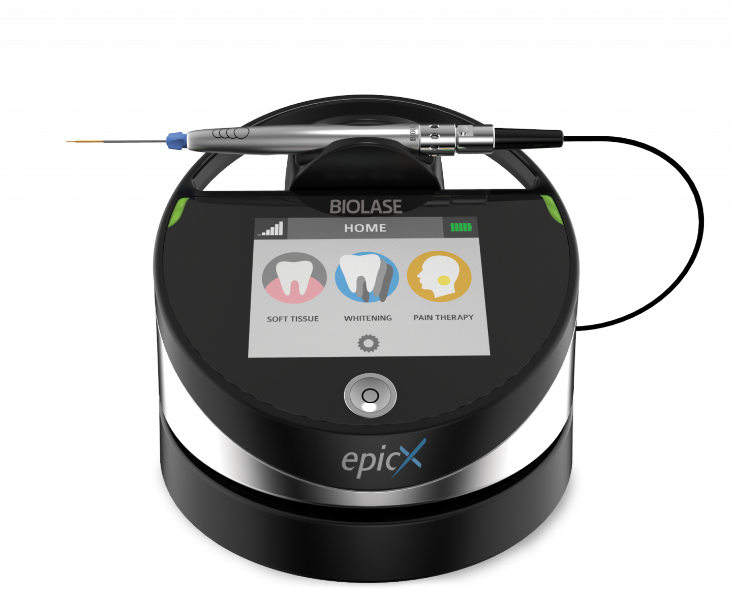
| Abstract: Aim The aim of the study was to accurately assess the antibacterial effect of the combined Er,Cr:YSGG and InGaAsP 940 nm laser therapy on nine pathogenic bacteria in the treatment of periodontitis. Materials and method Fifty-six patients were selected for this pilot study. Five patients were excluded, whereas 51 of them completed the study. The patients were randomly allocated to either the combined 2780 nm Er,Cr:YSGG (Waterlase, Biolase) and 940 nm InGaAsP diode laser (EPIC, Biolase) therapy, adjunct to scaling and root planning (SRP) (experimental group), or to scaling and root planning alone (control group). The quantitative and qualitative analysis of the total number of bacteria and nine specific germs was performed using quantitative real-time polymerase chain reaction. Results The total bacterial load inside the periodontal pockets was reduced both for the laser plus SRP and for the SRP alone group at the 1-month and 6-month follow-ups (p < 0.05). The laser therapy group showed a more significant bacterial reduction than the control group at the 1-month and 6-month follow-ups. The germ number reduction was statistically strongly significant for the total number of germs and for eight out of nine analyzed bacteria. Conclusions The present study suggests that a combined Er,Cr:YSGG2780 nm and diode InGaAsP 940 nm laser therapy added to the nonsurgical periodontal treatment brings an important benefit in bacterial reduction and stands as a reliable alternative to antibiotic prescriptions in periodontal treatment. The positive changes are also reflected in significant improvement of clinical periodontal parameters. The results suggest that newly formed bacterial microbiome inside the sulcus appears to be more beneficial, durable, and stable in the lased group. | ||||||
| Perio | DOI | Cobb, C.M., Low, S.B. and Coluzzi, D.J. | Lasers and the Treatment of Chronic Periodontitis [Abstract] |
Dental Clinics of North America Vol. 54(1), pp. 35-53 |
2010 | rank4 |
| Abstract: For many intraoral soft-tissue surgical procedures the laser has become a desirable and dependable alternative to traditional scalpel surgery. However, the use of dental lasers in periodontal therapy is controversial. This article presents the current peer-reviewed evidence on the use of dental lasers for the treatment of chronic periodontitis. textcopyright 2010 Elsevier Inc. All rights reserved. | ||||||
| Repair Perio | DOI URL | Dederich, D.N. | Periodontal Bone Regeneration and the Er,Cr:YSGG Laser: A Case Report. [Abstract] |
The open dentistry journal Vol. 7, pp. 16-9 |
2013 | rank1 |
| Abstract: BACKGROUND: Traditional methods of regenerating bone in periodontal bone defects have been partially successful and have involved numerous protocols and materials. More recently, it has been proposed that Er,Cr:YSGG laser energy may also be beneficial in the treatment of periodontal pockets, particularly in the regeneration of bone lost due to periodontal disease.nnCASE DESCRIPTION: The purpose of this paper is to present a case report of the Er,Cr:YSGG laser being used to conservatively treat a recalcitrant periodontal pocket in the presence of a periodontal bone defect and that resulted in successful resolution of the pocket and significant radiographic bone fill at the 1 year recall visit.nnCLINICAL IMPLICATIONS: This protocol using the Er,Cr:YSGG laser for the treatment of periodontal loss of attachment and periodontal bone loss may represent a less invasive alternative than traditional open-flap periodontal surgery or the intrasulcular use of other more penetrating laser wavelengths. | ||||||
| Perio | DOI URL | Dereci, Ö., Hatipoğlu, M., Sindel, A., Tozoğlu, S. and Üstün, K. | The efficacy of Er,Cr:YSGG laser supported periodontal therapy on the reduction of peridodontal disease related oral malodor: a randomized clinical study [Abstract] |
Head & Face Medicine Vol. 12(20), pp. 1-7 |
2016 | rank5 |
| Abstract: Background: This study aims to evaluate the efficacy of Er,Cr:YSGG laser assisted periodontal therapy on the reduction of oral malodor and periodontal disease. Methods: Sixty patients with chronic periodontitis were included in the study and allocated into two groups each containing 30 patients. The study was planned in a double blind fashion. Conventional periodontal therapy was performed in group 1 and conventional periodontal therapy was performed in association with Er,Cr:YSGG application in group 2. Periodontal parameters of probing depth, clinical attachment level, plaque index and bleeding on probing were measured with a periodontal probe. Quantitative analysis of volatile sulphure compunds (VSCs) were measured with a calibrated halimeter at baseline level and at post-treatment 1st, 3rd and 6th months. P values <0.05 were accepted as statistically significant. Results: There was a statistical significant reduction in VSC values in group 2 at post-treatment 3rd and 6th months (p < 0.05). Pocket depth values at post-treatment 1st month and bleeding on probing values at post-treatment 3rd and 6th months were significantly decreased in group 2 (p < 0.05). Intragroup statistical analysis revealed that there were statistically significant differences for all parameters (p <0.01). Conclusions: Er,Cr:YSGG laser assisted conventional periodontal therapy is more effective in reducing oral malodor and improving periodontal healing compared to conventional periodontal therapy alone | ||||||
| Repair Perio | URL | Dyer, B. and Sung, E.C. | Minimally Invasive Periodontal Treatment Using the Er,Cr:YSGG Laser [Abstract] |
The Open Dentistry Journal Vol. 6, pp. 74-78 |
2012 | rank4 |
| Abstract: Minimally invasive surgery (MIS) using the erbium, chromium: yttrium-scandium-gallium-garnet (Er,Cr:YSGG) laser (Waterlase MD, Biolase, Irvine, CA) to treat moderate to advanced periodontal disease is presented as an alternative to conventional therapies. To date, there are few short- or long-term studies to demonstrate the effects of this laser in treating and maintaining periodontal health. Electronic clinical records from 16 patients – total of 126 teeth, with pocket depths ranging from 4 mm to 9 mm – were treated with the same protocol using the Er,Cr:YSGG laser. The mean baseline probing depths (PD) were 5 mm and clinical attachment levels (CAL) were 5 mm in the 4 - 6 mm pretreated laser group. The mean baseline probing depths were 7.5 and 7.6 mm for PD and CAL respectfully in the 7 – 9 mm pretreatment laser group. At the 2 year mark, the average PD was 3.2 ± 1.1 mm for the 4-6 mm pocket group and the 7-9 mm pocket group had a mean PD of 3.7 ± 1.2 mm. mean CAL was 3.1 ± 1.1 mm for the 4-6 mm group and 3.6 ± 1.2 for the 7-9 mm group with an overall reduction of 1.9 mm and 4.0 mm respectively. At one and two years, both groups remained stable with PD comparable to the three-month gains. The CAL measurements at one and two years were also comparable to the three-month gains. | ||||||
| Perio | DOI URL | Ertugrul, A.S., Tekin, Y. and Talmac, A.C. | Comparing the efficiency of Er,Cr:YSGG laser and diode laser on human β-defensin-1 and IL-1β levels during the treatment of generalized aggressive periodontitis and chronic periodontitis [Abstract] |
Journal of Cosmetic and Laser Therapy Vol. 19(7), pp. 409-417 |
2017 | rank5 |
| Abstract: BACKGROUND The aim of this study is to determine the suitability of the Er,Cr:YSGG and 940 ± 15-nm diode laser for the treatment of generalized aggressive periodontitis and chronic periodontitis by measuring the levels of human β-defensin-1 and IL-1β. PATIENTS AND METHODS A total of 26 patients were included in this study. The study was designed as a "split-mouth" experiment. We performed scaling and root planing in the right maxillary quadrant, scaling and root planning + Er,Cr:YSGG laser in the left maxillary quadrant, scaling and root planning + 940 ± 15-nm diode laser in the left mandibular quadrant, and only scaling and root planing in the right mandibular quadrant. The presence of human β-defensin-1 and IL-1β was analyzed with an ELISA. RESULTS When the baseline and post-treatment human β-defensin-1 levels and IL-1β levels of the study groups were evaluated, a decrease in human β-defensin-1 and IL-1β were observed in the quadrant where the Er,Cr:YSGG laser was applied in both the generalized aggressive periodontitis group and the chronic periodontitis group. CONCLUSIONS The use of the Er,Cr:YSGG laser at non-surgical periodontal treatment decreased both IL-1β and human β-defensin-1 levels. It is likely that Er,Cr:YSGG laser is more suitable for the treatment of generalized aggressive periodontitis and chronic periodontitis. | ||||||
| Perio | URL | Ezzat, A., Maden, I., Hilgers, R.-d. and Gutknecht, N. | In vitro study: conventional vs. laser (Er,Cr:YSGG) subgingival scaling and root planing; morphologic analysis and efficiency of calculus removal using macroscopic, SEM and laser scanning [Abstract] |
Lasers in Dental Science Vol. February, pp. 1-7 |
2018 | rank3 |
| Abstract: Manyways and techniques were used for subgingival calculus removal to be achieved, fromscaling and root planning closed and opened technique, manual curettes, air abrasives, and finally dental lasers machines. Material and methods In this study, we will compare conventional ultra-sonic scaling and curettes to two laser groups using Er,Cr:YSGG 2780 nm. One group (LASER) using 25 mJ, 50 Hz, 50 μs pulse duration, 20 ms pulse period, 1.25 Waverage power, 500 W peak power W/A:80%/10%, and other group (LASER+) using manufacturer instructions: 47 mJ, 75 Hz, 50 μs pulse duration, 3.5Waverage power, 940Wpeak power, W/A40%, 20%. Fifteen extracted teeth that have subgingival calculus was usedineachgroup. Results Showed no statistical difference between time in three groups, there was a statistical difference concerning the amount of calculus, group laser + showed least amount of calculus remaining. Root roughness showed statistical significant, the laser + treatment shows significant higher root values than the laser treatment as well as compared to conventional treatment. Laser scanning showed no statistical difference concerning root roughness. SEM showed con- ventional group with roughness and smear layer, laser group showed the most smooth surface, laser + showed the most rough surface. Conclusion Er,Cr:YSGGis effective and efficient to remove subgingival calculus and safe to the pulp specially the parameters of laser group, as well as it is benefits concerning smear layer removal, bactericidal effect, promoting fibroblasts attachment and mostly few raise in pulpal temperature as shown in other studies. Amount of water spray is considered to be the most important factor due to it is effect on roughness and root morphology. Keywords | ||||||
| Perio | DOI | Fekrazad, R., Lotfi, G., Harandi, M., Ayremlou, S. and Kalhori, K.A.M. | Evaluation of fibroblast attachment in root conditioning with Er, Cr:YSGG laser versus EDTA: A SEM study [Abstract] |
Microscopy Research and Technique Vol. 78(4), pp. 317-322 |
2015 | rank5 |
| Abstract: The regeneration of periodontal support is a main concern in periodontal ther- apy. This study aims to investigate the efficacy of Er, Cr:YSGG laser and EDTA based condition- ing in attachment of fibroblast on root surfaces. This in vitro study was conducted on 81 root plates (6mm34mm31 mm) prepared from 27 single-rooted human mature teeth. The samples were divided into three groups: (1) Er, Cr: YSGG laser conditioning with a G6 tip (2.78 mm, 0.75 W, pulse duration of 140 ms, repetition rate of 20 Hz) for 5–7 s; (2) EDTA conditioning (17%, pH: 8) for 1 min; and (3) the control group which were exposed neither to EDTA nor laser. The viabil- ity and proliferation rates assessments were performed using MTT assay on days 3 and 5. In addition, the level of cell attachment was studied using scanning electron microscopy. The data indicated Er, Cr:YSGG conditioning increased cell viability by lapse of time (from days 3–5), with significantly better cell attachment compared to the other groups on days 3 and 5 (P<0.05). In addition, increasing cell attachment in the EDTA conditioning group compared with the con- trol group was statistically significant on day 5 but not on day 3 (P<0.05). In conclusion, Er, Cr:YSGG laser conditioning can promote enhance fibroblast attachment on dentinal root surfa- cesmore than EDTA. | ||||||
| Perio | DOI URL | Ge, L., Zhang, Y. and Shu, R. | Er,Cr:YSGG Laser Application for the Treatment of Periodontal Furcation Involvements [Abstract] |
Photomedicine and Laser Surgery Vol. XX(Xx), pp. 1-6 |
2016 | rank5 |
| Abstract: Objective: The purpose of this study was to evaluate the clinical use of an erbium, chromium: yttrium– scandium–gallium–garnet (Er,Cr:YSGG) laser in the management of root furcation involvements. Background data: It has been suggested that the use of laser radiation within the periodontal pocket may be effective in periodontal treatment. However, very limited data from clinical trials exist directly comparing the use of the Er,Cr:YSGG laser to conventional root planing. Materials and methods: Thirty patients with chronic period- ontitis were recruited. A total of 128 teeth with degree II or III furcation involvement were included in a split- mouth design such that the teeth on one side of the mouth were established as Group A and treated with the Er,Cr:YSGG laser and the teeth on the other side were established as Group B and treated with manual subgingival debridement. Changes in probing depth (PD), bleeding on probing (BOP), clinical attachment loss (CAL), and visual analogue scale (VAS) pain scores were compared between Groups A and B and within individuals over time at 6 and 12 weeks after treatment, testing the null hypothesis that there would be no clinical difference between treatment modalities. Results: Both treatments significantly reduced the PD, CAL, and BOP in the diseased teeth with degree II or III furcation involvement. The reduction of PD and BOP at weeks 6 and 12 was significantly higher in Group A (laser treated) than in Group B. The VAS pain score was significantly lower in Group A than in Group B ( p< 0.01). Conclusions: Pocket debridement with Er,Cr:YSGG laser is a safe, comfortable, and clinically effective means of subgingival debridement in periodontal therapy. | ||||||
| Open Flap, Repair Perio | DOI | Gupta, M., Lamba, A.K., Verma, M., Faraz, F., Tandon, S., Chawla, K. and Koli, D.K. | Comparison of periodontal open flap debridement versus closed debridement with Er,Cr:YSGG laser [Abstract] |
Australian Dental Journal Vol. 58(1), pp. 41-49 |
2013 | rank5 |
| Abstract: BACKGROUND: Traditional periodontal open flap debridement (OFD) results in reduced pocket depth (PD), clinical attachment loss (CAL), gingival recession (GR) and postoperative pain and discomfort. The quest to overcome these shortcomings has led to research into Er,Cr:YSGG laser assisted pocket therapy (ELAPT). This study was designed to compare the clinical outcomes of ELAPT versus OFD.n Fifteen patients with a PD of ≥5 mm and ≤8 mm at two sites were selected. Test sites (Group 1) were treated by ELAPT and the control (Group 2) by OFD. Clinical parameters were recorded at baseline, 3 and 6 months and included Plaque Index (PI), Gingival Index (GI), modified Sulcular Bleeding Index (mSBI), PD, CAL and GR.n Both treatments produced a reduction in PI, GI, mSBI and PD, an increase in GR, and a gain in CAL at 3 and 6 months. The mean gain of CAL in Group 1 at 3 and 6 months (1.60 ± 0.78 and 1.80 ± 0.63) was similar (p > 0.05) to the value of Group 2 (1.93 ± 0.88 and 2.00 ± 0.54). GR increased significantly (p < 0.05) only in Group 2 at 3 and 6 months (1.80 ± 0.56 and 1.87 ± 0.64) compared to Group 1 (0.50 ± 0.68 and 0.60 ± 0.74).n ELAPT compared with OFD results in similar CAL gains with less GR and significant reductions in PD, GI and mSBI, and may be considered as an alternative to surgical therapy. | ||||||
| Perio | DOI URL | Gutknecht, N., Van Betteray, C., Ozturan, S., Vanweersch, L. and Franzen, R. | Laser Supported Reduction of Specific Microorganisms in the Periodontal Pocket with the Aid of an Er,Cr:YSGG Laser: A Pilot Study [Abstract] |
The Scientific World Journal Vol. 2015(i), pp. 1-7 |
2015 | rank5 |
| Abstract: OBJECTIVE The aim of this study was to evaluate the effectiveness of a radial firing tip of an Er,Cr:YSGG laser as an adjunct to a nonsurgical periodontal treatment. METHODS Twelve patients with chronic or aggressive periodontitis were treated by conventional periodontal treatment using ultrasonic devices and hand instruments and, additionally, in two quadrants with an Er,Cr:YSGG laser. A new radial firing tip (RFPT 14-5, Biolase) was used with 1.5 W, 30 Hz, 11% air, 20% water, and pulse duration 140 μs. Microbiological smears were taken before treatment, one day after lasing, and three and six months after lasing. Pocket depths of all periodontal sites were measured before and six months after treatment. RESULTS The total bacterial load of Prevotella intermedia, Tannerella forsythia, Treponema denticola, Fusobacterium nucleatum, Porphyromonas gingivalis, and Aggregatibacter actinomycetemcomitans inside the pocket was reduced significantly throughout the whole examination time. Greater pocket depth reductions were observed in all groups. There was a slight higher reduction of pocket depth in the lased group after six months. CONCLUSIONS These results support the thesis that Er,Cr:YSGG laser supported periodontal treatment leads to a significant reduction of periopathogenes and thereby helps the maintenance of periodontal health. | ||||||
| Perio | DOI | Hakki, S.S., Berk, G., Dundar, N., Saglam, M. and Berk, N. | Effects of root planing procedures with hand instrument or erbium, chromium:yttrium-scandium-gallium-garnet laser irradiation on the root surfaces: A comparative scanning electron microscopy study [Abstract] |
Lasers in Medical Science Vol. 25(3), pp. 345-353 |
2010 | rank5 |
| Abstract: The purpose of this study was to investigate the efficiency of hand instrumentation and laser irradiation on calculus removal from the root surfaces, in vitro. Thirty-two human teeth, extracted for periodontal reasons, were used in this study. Root surfaces of single-rooted teeth were treated by different methods including (1) conventional hand instruments; (2) hand instruments and tetracycline-hydrochloride (Tet-HCl); (3) erbium, chromium:yttrium-scandium-gallium-garnet (Er,Cr:YSGG) laser irradiation, setting I (short pulse); (4) Er,Cr:YSGG laser irradiation, setting II (long pulse). Three premolar teeth, extracted for orthodontic reasons, served as control. The morphology of the root surfaces was evaluated by light and scanning electron microscopy. Energy dispersive X-ray (EDX) analysis was performed to compare the mineral content of root surfaces treated with hand instrumentation and lasing procedures. The results of this study demonstrated that all treatments were efficient in calculus removal from the root surfaces. Thermal changes, including melting and carbonization, were not observed in either lasing procedure. The surface was rougher in the laser groups than in the groups treated with hand instruments. Moreover, roughness was greater in the long-pulse laser setting than in the short-pulse setting. While increased calcium (Ca) and decreased phosphate (P) (weight concentration percent) were observed in all treatments when compared with the control, laser procedures resulted in a more similar mineral content than in the groups treated with hand instruments. Based on these findings, laser procedures, when used in appropriate settings, are capable of performing scaling and root planing in the treatment of periodontitis. It may be concluded that short pulse laser may be more suitable for the micro-morphology of the root surface. However, additional in vitro and clinical studies are necessary to clarify the success of laser in periodontal therapy. | ||||||
| Perio | DOI | Hakki, S.S., Korkusuz, P., Berk, G., Dundar, N., Saglam, M., Bozkurt, B. and Purali, N. | Comparison of Er,Cr:YSGG laser and hand instrumentation on the attachment of periodontal ligament fibroblasts to periodontally diseased root surfaces: an in vitro study. [Abstract] |
The Journal of periodontology Vol. 81(8), pp. 1216-1225 |
2010 | rank5 |
| Abstract: This study investigates the effects of erbium, chromium:yttrium-scandium-gallium-garnet (Er,Cr:YSGG) laser irradiation and hand instrumentation on the attachment of periodontal ligament (PDL) fibroblasts to periodontally involved root surfaces. | ||||||
| Repair Perio | DOI | Kelbauskiene, S., Baseviciene, N., Goharkhay, K., Moritz, A. and Machiulskiene, V. | One-year clinical results of Er,Cr:YSGG laser application in addition to scaling and root planing in patients with early to moderate periodontitis [Abstract] |
Lasers in Medical Science Vol. 26(4), pp. 445-452 |
2011 | rank5 |
| Abstract: In 30 patients with periodontitis, a total of 278 teeth exhibiting bleeding on probing, subgingival calculus, and a probing depth between 3-6 mm were examined. For each participant, two treatment types were alternatively applied on the contralateral quadrants: scaling and root planing (SRP) as control, and SRP followed by Er,Cr:YSGG laser application (SRP+laser), as a test method. Five clinical parameters: plaque level, bleeding on probing, probing depth, gingival recession and clinical attachment level were examined at baseline and at 2, 3, 6, 12 months after treatment. Of the total of 1,668 sites examined in all patients, 1,088 sites were found with a probing depth of 3-6 mm. In these sites, differences in clinical parameters between SRP and SRP+laser-treated quadrants were analyzed, assuming the level of p < 0.05 as significant. After 2 months from baseline, the mean probing depth reduction and the clinical attachment level gain were significantly greater in SRP+laser than in SRP quadrants, and remained so throughout the study (p < 0.001). A marked reduction of the bleeding scores occurred in all examined sites, irrespective of the treatment method. However, after 12 months, significantly less teeth exhibited bleeding on probing in SRP+laser quadrants than in SRP quadrants (p < 0.001). The mean plaque and gingival recession levels did not differ between the SRP and SRP+laser quadrants neither before nor after the treatment. The periodontal procedures either using Er,Cr:YSGG laser after SRP or SRP alone, lead to significant improvements in all clinical parameters investigated. However, laser application, as an adjunct to SRP, appeared to be more advantageous. | ||||||
| Perio | URL | Miteva, M., Peev, S. and Hristov, I. | The Adjunctive Use of the Er , Cr : YSGG Laser in Nonsurgical Periodontal Treatment [Abstract] |
International Journal of Science and Research, pp. 61-64 | 2017 | rank4 |
| Abstract: The aim of this study was to evaluate the effectiveness of a radial firing tip of an Er,Cr:YSGG laser as an adjunct to a nonsurgical periodontal treatment. Fifteen patients with chronic periodontitis were treated by conventional periodontal treatment using ultrasonic devices and hand instruments and fifteen patients with additional use of Er,Cr:YSGG laser. A radial firing tip (RFPT 14-5, Biolase) was used with 1.5 W, 30 Hz, 11% air, 20% water, and pulse duration 140 μs. Probing pocket depths, gingival margin and clinical attachment level were taken before treatment, a month and a half and three months after lasing. Both treatment modalities were effective in treating chronic periodontitis. Keywords: | ||||||
| Perio | DOI | Noori, Z.T., Fekrazad, R., Eslami, B., Etemadi, A., Khosravi, S. and Mir, M. | Comparing the effects of root surface scaling with ultrasound instruments and Er,Cr:YSGG laser [Abstract] |
Lasers in Medical Science Vol. 23(3), pp. 283-287 |
2008 | rank4 |
| Abstract: There are several studies done to show the comparability of laser scaling and root planing with routine methods. The most suitable wavelengths for such an application are reported as 2,940 and 2,780 nm. The superficial interactions of the current wavelength with root surface is investigated in this study to compare the crater and crack formation during the procedures between ultrasound and Er,Cr:yttrium-scandium-gallium garnet (YSGG; 2,780 nm) laser-treated teeth. Thirty human teeth with calculus on their root surface, which were extracted because of the severe periodontitis, were selected for this interventional in vitro study. Calculus area were divided into two equal parts: One of them was prepared for Er,Cr:YSGG (Biolase, Waterlase, USA) laser irradiation and the other one for ultrasound treatment (Dentsply cavitron, DENTSPLY International, USA). The Er,Cr:YSGG laser was specified as follows: pulse energy = 50 mJ, power = 1 W, wavelength = 2,780 nm, pulse repetition rate = 20 pulse per seconds, tip length = 6 mm, and tip diameter = 600 microm. Nineteen of the laser samples (95%) and eight of ultrasound ones (40%) out of 20 samples in each group had craters showing a statistical significant difference (p < 0.001). The number and depth of these craters are also evaluated. Thirteen samples of the laser cases (65%) and all samples of the ultrasound group showed cracks with significant differences (p = 0.008). In addition, the number and width of cracks in both groups are reported. As a general conclusion, the laser-treated samples show more craters but less cracks. | ||||||
| Perio | DOI URL | Ting, C.-C., Fukuda, M., Watanabe, T., Aoki, T., Sanaoka, A. and Noguchi, T. | Effects of Er,Cr:YSGG laser irradiation on the root surface: morphologic analysis and efficiency of calculus removal. [Abstract] |
Journal of periodontology Vol. 78(11), pp. 2156-64 |
2007 | rank5 |
| Abstract: This in vitro study was performed to determine the appropriate power output setting for an erbium, chromium-doped:yttrium, scandium, gallium, and garnet (Er,Cr:YSGG) laser used in periodontal pocket irradiation by examining the morphologic alterations of the root surfaces and the efficiency of calculus removal. | ||||||


